High-throughput/high-content virology and screening
In the past decade, high-throughput screening has become a critical tool in antiviral discovery. More recently, advances in high-throughput and high-content imaging have further advanced unbiased phenotypic screenings, allowing to quantitate subcellular events beyond traditional cytopathic effects. At VRS we have used both GFP-tagged viruses and immunofluorescence staining to precisely assess the ability of compound libraries to inhibit virus infection, in 96 and 384 well plate format. Depending on the antigen that we chose to visualize, we have been able not only to measure percentages of infection, but also to observed localization of viral proteins and replication complexes, or even patterns of virus transmission.
High-throughput, high relevance
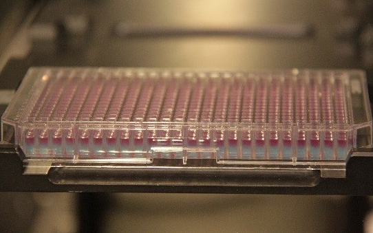
The advantages of high-throughput imaging go beyond testing large numbers of compounds. Running informative experiments in 96 or 384 well format greatly enhances the capability of any assay, as it allows parallel testing of multiple conditions, introduction of multiple controls, higher number of replicates, and use of minimum amounts of precious samples. This translated in more controlled and more robust results, as well as the opportunity to visually assess the information acquired. Also, some of the traditional virology assays, like plaque assays or immunofocus assays, due to the larger well size can use up a lot of sample very quickly. By using high-throughput imaging, our clients have been able to use the same 200 μl of sample for triplicate dose-response testing against over five viruses (we could have carried on, but they only needed those five..).
If you can see it you can measure it!
Better microscopes and acquisition software are not the only advances in high-throughput imaging: an incredible leap forward has been made on the analysis softwares. New programs are provided with sophisticated algorithms that batch process thousands of pictures automatically and are able to quantify a large variety of subcellular phenomena, including punctate objects, re-localization events, or alterations in organelles morphogenesis on a single cell basis. Assays that would have involved hours on a confocal microscope to acquire a representative number of cells and an equal amount of time (and computer crashing!) for processing, can now be performed semi-automatically, with the possibility of manually inspect the process at any point. Using the Perkin Elmer Opera and Opera Phenix systems and the associated Columbus and Harmony software, at VRS we have been able to quantify and correlate mean fluorescence intensities, fusion events, and internalization of viruses, molecules, and dyes. Interestingly, robust and reproducible data have been obtained even for phenomena that were unclear by eye, revealing information that otherwise may have been missed.
If your research project involves immunofluorescence, or if you want to maximize the information generated from your experiments, contact us to find if you could benefit from high-throughput and high-content imaging.