Understanding at what stage of a virus life cycle a compound or a restriction factor acts is critical in the discovery process. In drug discovery, this aspect can be a bottleneck for hits generated via phenotypic screenings, but even when the viral of cellular targets are known it is important to confirm that a given molecule does indeed act where it is supposed to.
In this series of three blogs we present different assays and techniques that can be used to narrow down the mode of action of compounds or cellular proteins that interfere with the virus life cycle, and we focus on procedures that – with some optimization – can by applied to different viruses. In this first blog we cover assays that explore the early stages of viral entry and fusion; in the next we will look at viral replication; and in the third we will follow assembly, release, and spread. Ready to follow where your virus gets stuck?
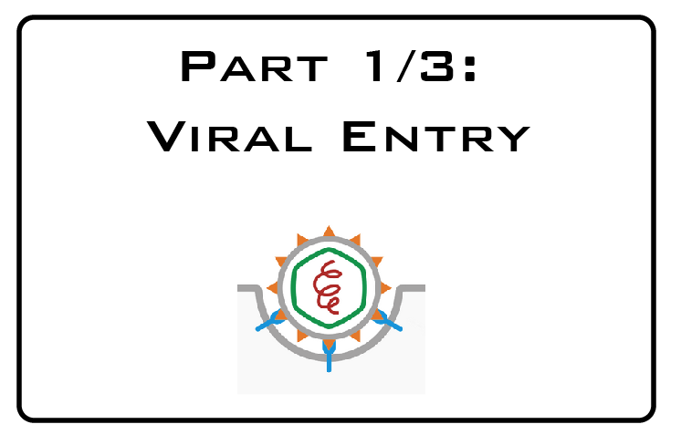
Part 1/3: Viral entry
Although nature has devised a lot of strategies to inhibit virus infection at different stages, it has put considerable effort in blocking viral entry. Stopping a virus on its way in is what antibodies do. More recently, a class of proteins called InterFeron Inducible TransMembrane (IFITM) proteins has been shown to effectively interfere with fusion of a wide variety of viruses.
Blocking entry is powerful because, by preventing infection all together, it saves a lot of troubles from the start. Also, a virus that hasn’t been given the chance to enter the cell and replicate is very unlikely to be able to develop resistance against that early block.
Different viruses, similar strategies
While different viruses can use different entry pathways (like fusion at the plasma membrane or endocytosis, pH-dependent or pH-independent fusion), the general principles appear to be conserved: every virus needs to 1) bind to the cell membrane, 2) be internalized, and 3) release its genetic information into the cytoplasm or the nucleus. While specific assays exist for individual viruses, these general principles mean that several experimental procedures are amenable to study the mode of action of antiviral compounds, cellular restriction factors, or antibodies against many different viruses.
Does an antibody prevent virus internalization, or does it allow internalization but then block the next critical step of virus fusion that allows genome release in the cytoplasm? This can be an important question. Think of viruses like dengue, where the antibodies generated by a primary infection can enhance secondary infections by helping the virus in through binding of the Fc receptor: if these antibodies could still block fusion, virus internalization may be less of a problem.
Studying entry: the toolbox
Virus entry can be study using microscopy, biochemical assays, or some clever enzymatic reaction upon tagging of virus components.
Electron microscopy (EM) has been paramount to understand many details of viral entry, as it can clearly see viral particles smaller than 50nm inside endosomes. The problem with EM is that it requires large amounts of virus, patience, and highly skillful expertise. If you have all of this, the results are highly gratifying!
Recent advances in fluorescence microscopy have also proven invaluable for the study of viral infection and particularly entry. Larger and robust viruses tolerate the presence of even relatively large fluorescent tags (like GFP) on their structural proteins: live imaging than allows following these viruses all the way into the cells, while labelling of cellular markers can additionally help localize the virus in subcellular organelles. Much smaller tags have also been developed that are not themselves fluorescent, but bind to fluorescent dyes separately added in the media. These are generally tolerated also by smaller and unstable genomes, helping the visualization of a wider array of viruses.
Highly specific antibodies are now available for many viruses. Antibody staining generally requires fixation, but allows visualization of wild type viruses at the desired time point, making it possible to distinguish, for instance, between virus bound to the cell surface and internalized virus. When good antibodies are not available, fluorescence in situ hybridization (FISH) can be a solution. FISH uses fluorescent probes complementary to the viral genome to allow visualization of either the incoming virus or replicating virus. Probes can be designed or purchased, providing a flexible approach.
Biochemistry techniques tend to rely on radio-labelled virus, biochemical reactions, or western blots. The first is a highly sensitive technique, which is however more rarely adopted these days due to the associated health & safety risks. The last is far less sensitive and generally requires higher amount of virus, as well as good antibodies, but is often much easier to implement.
Did it bind to the cell?
Cell adhesion and receptor binding are the first steps of virus infection. By incubating virus and cell at 4°C for >30 minutes, binding can be synchronized but internalization or fusion are prevented. If the experiment is stopped at this point, either with fixation (for microscopy) or cell lysis (for biochemical assay), it is possible to determine if a certain treatment has reduced the amount of virus bound to the cell surface compared to control. If a fixative is used for this experiment, it is important that it is free of methanol, as this can introduce pores on the membrane confounding the results.
Was it internalized?
After synchronizing the binding at 4°C, virus internalization can be induced by warming the culture to 37°C. Internalization tends to happen fairly instantly, while fusion is better seen after 15-20 minutes, however the exact timing for each virus will need to be determined experimentally. Again, microscopy will allow visualization of a virus inside the cell, or even localized in specific compartments that have been tagged or immune-labelled for ease of identification. If biochemistry is the method of choice, one cleaver trick is to stop the downstream events by moving the culture back on ice, and cleave any virus that has not been internalized with a protease like trypsin or subtilisin: if virus signal can still be detected after treatment this suggests that it has been internalized and is protected from the protease at the cell surface. Which protease, inhibitor, and kinetics will all have to be determined experimentally.
Did it fuse?
Fusion is the most critical stage of virus entry. If a virus fails to fuse and to release its genome into the cytoplasm, it will not be able to replicate and eventually it will be destroyed by the cell. An antibody recognizing the virus capsid (rather than the envelope) can help visualize fusion by microscopy, however this may be less straightforward, reason for which more specific assays have been devised.
If the virus of interests tolerates it, the beta-lactamase enzyme can be tagged to a viral protein that is integral part of the genome. A fluorescent substrate is then added to the culture and internalized in the cell cytoplasm: this will become available for cleavage by the beta-lactamase enzyme only once this has also been released in the cytoplasm following a fusion event. This approach has been devised for instance for HIV, Ebola virus, and influenza, but is not tolerated by every virus. A more generic approach for enveloped virus involves the incorporation of lipophilic dyes (like DiD or R18) into the virus membrane, where they self-quench due to close proximity. Only upon fusion of the viral membrane with the cellular membrane, the dyes diffuse and emits signal in the far red.
Time is your friend
If you are working with compounds, the timing of administration will provide useful information on the infection stage inhibited. Pre-incubation of virus and compound can reveal whether the drug neutralizes the virus directly. Equally, addition of a drug before or at different times after infection can inform whether the compound is more likely to inhibit early or late stages. For all these assays we cannot emphasize enough the importance of optimization and especially controls. Drugs or proteins that are know to inhibit the stage of interest should be included to confirm assay specificity, as well as untreated and uninfected controls. Of course, you can leave the fuss to us and just enjoy knowing where your virus gets stuck!