Welcome to our blog about air-liquid interface (ALI) testing services. As the demand for more effective and reliable preclinical models continues to grow, the use of ALI technology has become increasingly popular. At Virology Research Services, we provide ALI testing services to help our clients make more informed decisions about the efficacy of their compounds.
In this blog, we will explore the benefits of ALI testing, the types of studies that can be performed using this technology, and how our services can help you achieve your research goals.
Cell culture in virology
Cell culture is one of the most important, accessible, and versatile tools in virology. Most cell culture work done in virus labs revolves around growing cells permissive to the virus of interest, then characterizing the virus and how it grows, or challenging the virus with various chemical or biological agents. We also use cell culture to grow virus stocks; the virus is allowed to replicate in cultured cells before collecting the resulting supernatant. These virus stocks can then be stored for further use.
The limitations of traditional culture methods
The monolayer culture system is a useful tool, but it does not accurately represent real-world multi-layered biological systems.
For example, the respiratory system is a multi-cellular environment that cannot be accurately replicated by growing just one cell type. The lungs and airways are complex organs spread over approximately 80 m2 and consist of goblet, basal, and ciliated epithelial cells, among other rarer cell types. In a healthy person, these cells work together to produce a healthy respiratory environment, with the goblet cells releasing the mucus that traps inhaled pollutants and pathogens and the ciliated epithelia sweeping this mucus up and out of the lungs. Clearly, a simple monolayer culture system cannot capture this complexity.
The growing importance of representative respiratory models
Unfortunately, of the millions of particles we inhale every hour, some – including respiratory viruses – will persist and cause damage, sometimes leading to sickness. Given the events of recent years, few of us need reminding of how respiratory viruses can lead to severe illness and death.
Introducing Air-Liquid Interface (ALI) culture
Until recently, cell culture was limited to growing a monolayer of these permissive cells in a plastic flask or dish. But to satisfy the demand for more suitable lab models of respiratory infection, research scientists have developed methods to simulate a portion of the respiratory tract in three-dimensional (3D) cell culture. This approach is called Air-Liquid Interface (ALI) culture. This culture method is named ALI because the cells sit on a membrane (interface) that has one side exposed to the external environment (air) and the other exposed to the growth media (liquid).
How ALI is established
The process begins with a source of cells – this can be directly taken from a patient/volunteer, a section of an organ (e.g., a lung biopsy), or commercially-available starter cells.
The first step is to expand the basal cells by seeding them onto a collagen-coated flat vessel. This takes approximately 1 week and allows any debris or non-epithelial cells to be removed by washing, while the basal cells remain tightly adhered to the collagen.
Once these cells have grown to cover the surface of the well, an enzyme is added that gently detaches the cells from the plastic, and cells can be moved into a larger vessel. During this step, cell numbers can increase exponentially over just a few days. Once this larger vessel is covered (confluent), we again detach and count the cells.
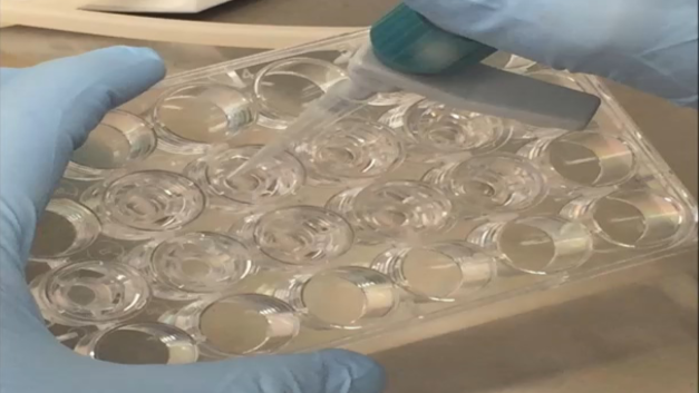
The next step is to induce these cells to differentiate into other cell types to become the representation of the lung environment we desire. To this end, cells are added to a Transwell, a small, porous vessel designed to sit on the well of a a plate so that the bottom is slightly above the surface of the well. This allows the specifically designed growth media to flow under the surface of the Transwell, bathing the cells from the bottom. This allows the cells to metabolize the nutrients in the media below, and continue their natural growth pattern and spread across the membrane until confluent. As they are still growing, we also continue to add growth media on top of the cells. Once they are at this confluent stage and the cells have formed tight junctions among each other, the next stage can start.
We now have a Transwell with a completely confluent sheet of basal epithelial cells, and we must give them the environment they need to start differentiating. The first step is to remove all the growth media from above and below the cells, and replace it with a differentiation media. From now on, no media is added to the top of the cells; so feeding is only provided through the bottom of the Transwell, through a porous membrane.

This differentiation media causes the basal cells to start producing other cell types on top of the basal cells to produce a multicell layer. These are the goblet, ciliated, and rarer cell types mentioned above, recreating the complexity of a respiratory multilayer. In the following days, the cells become elongated and columnar. The goblet cells start to produce mucus, and the cilia start to grow and eventually beat. If successful, a fully differentiated Transwell can be produced in 3–6 weeks after seeding.
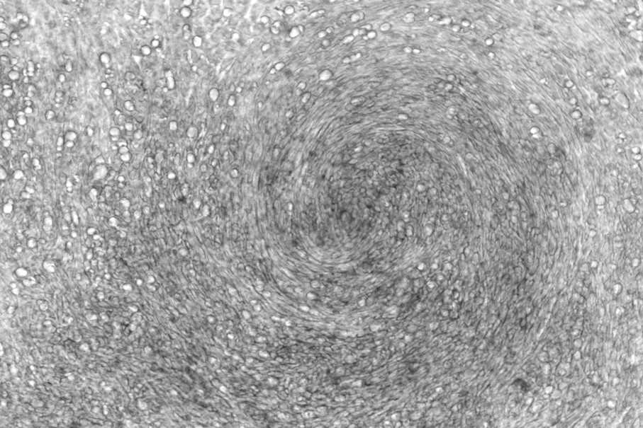
The cells are exposed and are differentiating, as can be seen from the morphology change from the “cobblestone” appearance to the spiralling elongated cells seen in the image centre.
At this point, cells behave as close to a sample directly from the respiratory system as possible, but without the problematic contamination associated with patient derived samples. These cells can then be used for in vitro studies, generating richer data than is possible with simple monolayer experiments.
Aspirating the mucus from the surface of the transwell before infection.
How the ALI system can be used
In the hands of experienced scientists, the ALI system can be a powerful tool with many applications. Example use cases for ALI include:
- To visualize the respiratory epithelium (biology and structure) before and after infection or a treatment.
- To study the infection process of the respiratory epithelium: For example, pathogens can be added to the fully differentiated cells to study their effects on cell death, cilia beat frequency, and mucus production.
- In respiratory disease models: 3D cell models can be used to study various diseases, such as asthma, cystic fibrosis, and chronic obstructive pulmonary disease. This can help describe the effects of known and novel drugs to treat these conditions.
- When testing novel drug formulations for inhaled delivery: Because ALI models are a relatively accurate representation of the in vivo situation, inhaled drugs can be deposited on the surface of ALI cells and monitored for uptake/effects, such as differences in transepithelial electrical resistance and pH.
- To test new technologies, such as nanoparticles and CRISPR.
- To test the toxicity of inhaled substances: ALI has shown to be a good representation of the human airway when subjected to inhaled toxic substances, such as air pollutants and tobacco smoke.
- To create complex co-culture system with immune cells to mimic the immune environment in the airways.
Our expert team can develop your ALI project
Here at Virology Research Services, we’re committed to providing high-quality in vitro testing to help our clients achieve their research goals. We offer a range of advanced testing methods, including the air-liquid interface (ALI) system.
Our ALI system is designed to mimic the environment of the lungs. With this advanced technology, our team of experts can help you design and conduct better in vitro studies.
At Virology Research Services, we have learnt to work with this powerful system to provide more physiologically relevant models of respiratory infections. At our state-of-the-art facility, we routinely produce high-quality ALI cultures which we then use for experiments and also make available to our clients.
As scientists, we understand the importance of flexibility and can offer ALI culture at different stages of differentiation and in different formats. We can then conduct the experiments in our labs, or ship the cultures directly to you for your end-use of choice. Our dedicated team of experts is committed to supporting your research and providing flexible solutions tailored to your specific requirements.
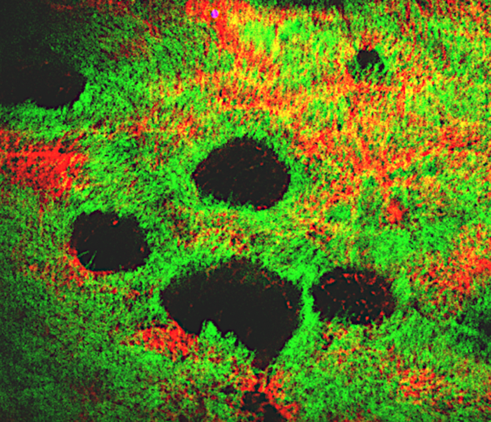
Confocal image of our differentiated ALI cells. The cilia are shown in green and actin in red.
Example Uses
Our ALI services are ideal for pharmaceutical companies developing new antiviral drugs for respiratory infections. By choosing our ALI service, it’s possible to leverage our expertise in growing, treating, and infecting ALI cultures with the virus of interest, as well as our ability to gather and analyse data.
Our ALI products are perfect for research labs that want to use a more physiological cell model in their research area and can use their in-house expertise and equipment for conducting their experiments. By opting for our ALI product, these labs can receive high-quality, ready-to-use ALI cultures without the need to establish and optimise the culturing process themselves.
By using our ALI services and products, researchers can save valuable time and resources, ensuring they can focus on their experiments and promptly advance their research.
Contact us today, and one of our scientists will get right back to you.
Blog by Paul Griffin
Edited by Reckon Better Scientific Service