Organoids
3D CELL MODEL
Bronchial organoids
Bronchial organoids are advanced 3D models that accurately mirror human airway tissue. Created from Normal Human Bronchial Epithelial (NHBE) cells, these structures replicate key features of airways including diverse cell types, tissue organization, mucus production, and ciliary movement. This model bridges traditional cell culture and animal testing, accelerating both our understanding of respiratory diseases and antiviral drug development.
Our Approach
How we grow organoids
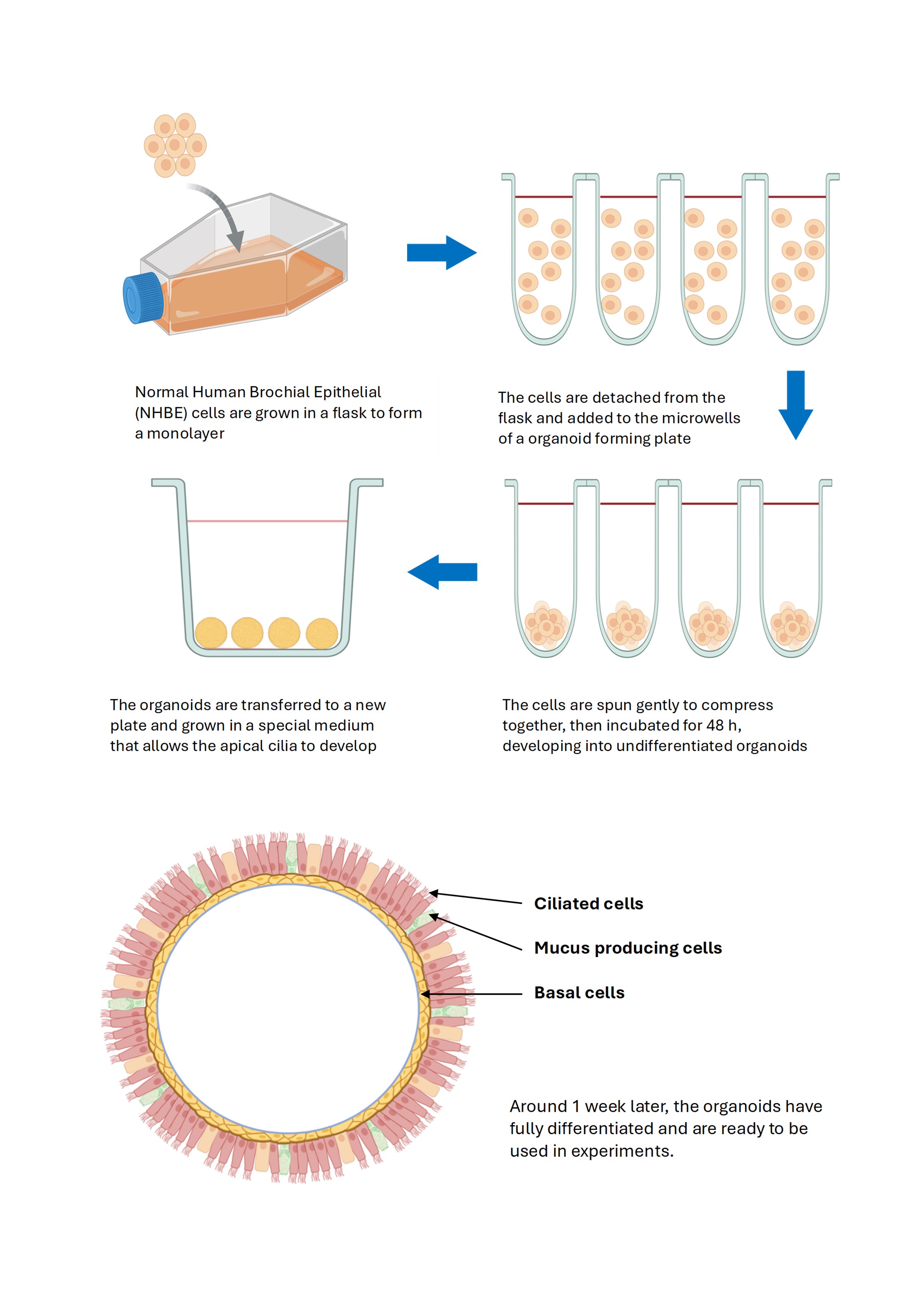
Replicates airway features: Our advanced organoid technology transforms normal human bronchial epithelial (NHBE) cells into fully-functional airway models. Starting with a monolayer culture, cells are carefully transferred to specialized microwells where they undergo controlled aggregation for 48 hours. The resulting undifferentiated organoids are then cultured in medium that promotes apical development.
After approximately one week, these organoids mature into complex structures featuring three key cell types found in human airways: ciliated cells for particle clearance, mucus-producing cells for protective barrier function, and basal cells that maintain tissue renewal. This physiologically-relevant model provides an ideal platform for respiratory disease research and drug development studies.

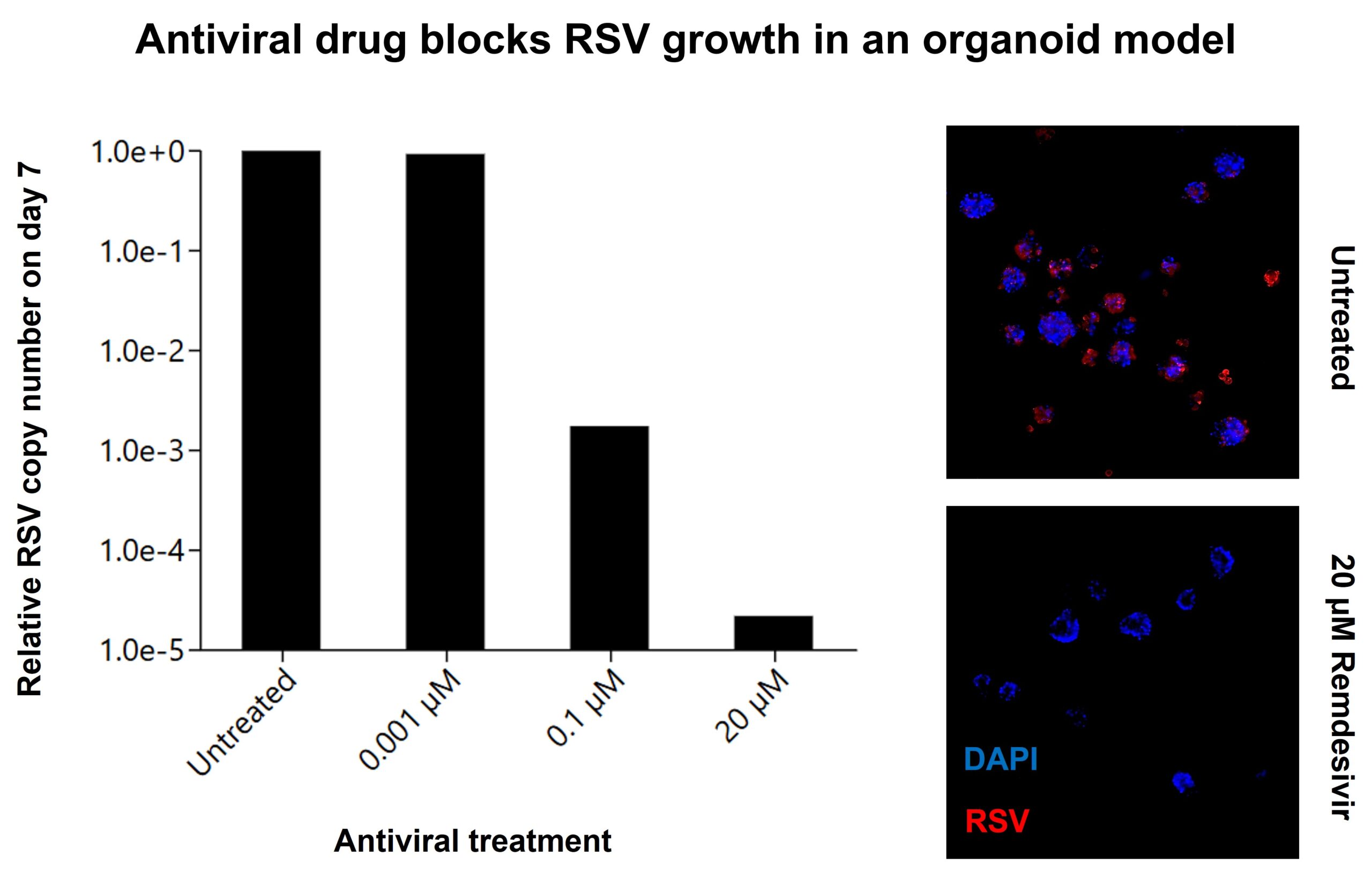
In this organoid 3D cell model case study, we detect a dose-dependent inhibition of RSV by the drug candidate in organoids, with a ~100,000-fold reduction at 20 µM. The microscopy images confirm this, showing abundant RSV (red) in untreated organoids but minimal infection in treated ones, while nuclei (blue) remain intact.
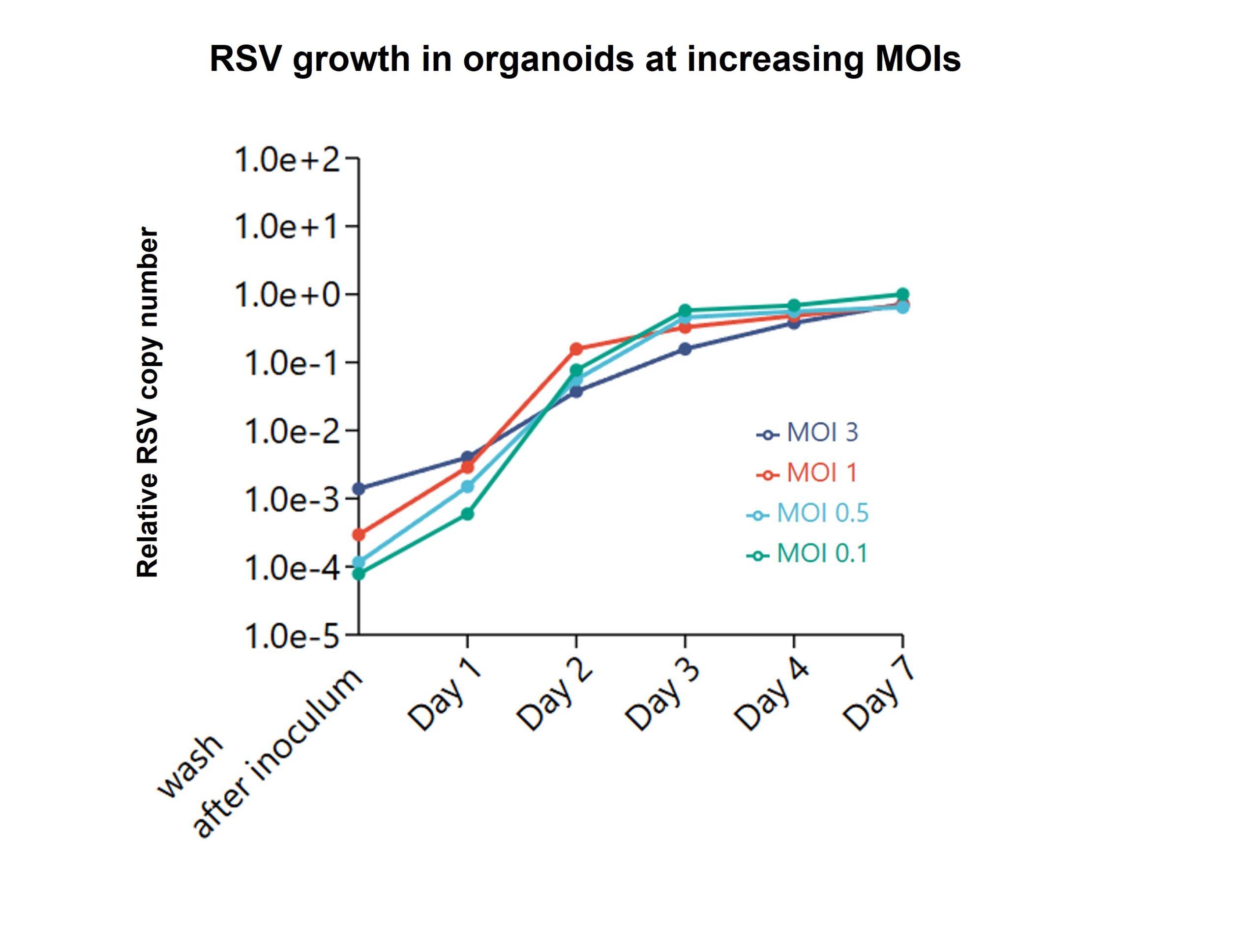
We tested RSV infection kinetics across multiple MOIs in bronchial organoids. Despite different initial virus loads, all MOIs reached similar RSV levels by day 7, with most viral growth occurring between days 1-3. The data shows MOI 0.1 is sufficient for maximum viral yield, enabling efficient virus usage in future experiments.


