This month we cover an old classic, the Tissue Culture Infectious Dose 50 assay, or TCID50. The TCID50 assay is used to quantify viral titres by determining the concentration at which 50% of the infected cells display cytopathic effect (CPE). As long as the virus of interest causes cell death, this assay has the advantage of being cheap and easy to implement also when virus-specific antibodies are not available. In fact, very little information on the virus itself is required, making it a key tool to study new and emerging pathogens.
TCID50 assay: what it is and how to use it
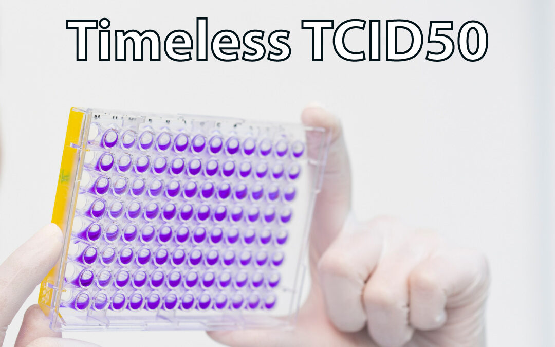
In a TCID50 assay, serial dilutions of a virus are added onto monolayers of cells, and left until CPE can be seen. At this point, the effect of the serial dilution on the cells can be scored either in a discontinuous or continuous manner. In a discontinuous manner, the single dilution at which 50% of cells display CPE is used to approximately quantify the amount of virus present in the original stock. This is fairly straightforward and doesn’t require further analysis. However, it also comes with a degree of inaccuracy, as it doesn’t allow to accurately distinguish within the same dilution.
More accurate information can be achieved in a continuous manner by using a colorimetric readout, e.g. a metabolic substrate with specific absorbance, where the amount of signal acquired is proportional to the number of live cells. The absorbance values of the different dilutions can be then plot in a continuous non-linear regression dose-response curve, from which it will be possible to extrapolated more accurate TC50 values. The discontinuous method is sufficient if a precise titre is not required, or to determine what concentration of virus is needed to obtain a certain percentage of infection in downstream assays (an advantage of the TCID50 is that it can be performed on the cell line of interest, as long as the virus causes CPE). However even in these cases it may be better to use dilutions folds between 1:2 and 1:5 to improve accuracy. When instead a more accurate quantification is necessary, for instance when comparing the effect of different treatments or mutations on virus titres, a colorimetric method may be preferred.
From dilutions to titres
TCID50 values give an indication of how many viruses is needed to have CPE in 50% of the cells. But how to go from this to the actual amount of virus per ml? The formula is quite simple, and it consists in multiplying the TCID50 value by 0.7. This comes from the Poisson distribution applied to viral infection which states that, in a fully permissive cell line, the probability of reaching 50% infection is achieved by a multiplicity of infection of approximately 0.7. This is not always true, but it’s a good approximation for most applications.
The troubles of counting viruses
As accurate as one can be, counting viruses is never easy. First, serial dilutions are -by their own nature- a source of error. Second -and this is particularly relevant for high titres of virus- even the tiniest volume that remains attached to the very end of a pipette tip can carry enough viral particles to make a substantial difference in the quantification. Third, the biological variation of the system is high. Plate the same amount of cells, add the same amount of virus, stop the infection at the same time, and the percentage of infection may be close, but never exactly the same.
Finally, when assessing a treatment that (as you would hope!) decreases virus titres, the amount of virus may fall below the assay detection threshold.
The art of counting viruses
The good news is that once you know your problems you are not far from a solution! Accuracy in serial dilution can be reduced by automation, but even when this is not available, some small tips can make the difference. These include changing pipette tips and never insert a pipette tip deeper than a millimetre into the next dilution, to avoid accidentally carrying over some extra logs of virus that just got stuck on the outer plastic. Replicates are also important, and the more so for miniaturised assays. 96 well plates allow simultaneous handling of multiple samples in a way that more low-throughput methods (like plaque assay) wouldn’t allow, but they come with additional variability, as small volumes are even more sensitive to the issues discussed above. While technical replicates go some way to address this, at VRS we try to run experiments that will require a colorimetric TCID50 in biological triplicate. This increases the robustness of the approach and allows to exclude spurious points in the dataset without losing significance. Accurate TC50 values will require a non-linear regression dose-response of the colorimetric data, which programmes like GraphPad Prism can easily generate. However, the ability of any software to plot this kind of curve will strictly depend on the quality of the data, and on the close numerical values of the replicates. A visual inspection of the curves and the TC50 values generated is highly recommended to correct situations where the extrapolation has been skewed by spurious data points. Above all, our experience suggests that beyond standard virus stock titrations, experiments that will require TCID50 readout may need additional steps of optimization, both to determine the best biological conditions that will assure upstream collection of reproducible amounts of virus (e.g. cell line of choice, plate format, multiplicity of infection, time points), and the best range of concentrations to test on the TCID50 assay (e.g. starting concentration and dilution fold).
Cell death or nice pictures?
Counting dead cells is not easy, as very often they abandon the plate at the first wash – supposing they are still there in the first place! This doesn’t help with accuracy. Although more expensive and laborious, whenever a good antibody is available, immunofluorescence staining is the approach that we would generally choose. The assay can be stopped before cell death and the sensitivity is generally higher, which allows more control and higher accuracy. Additionally, this method can be used for viruses that do not cause CPE, or that take a long time to do so (and lets face it, some nice pictures of a virus in action are far more uplifting!). Cell damage, however, is the trademark of most infections and of several emerging viruses of which we know very little. Several colorimetric/fluorescent reporters are now available to sensitively measure and distinguish between different types of cytotoxicity. It looks like this old classic is up for new challenges!