It’s 1907, and Ross G. Harrison has just developed a method of cultivating animal tissues outside of the body by removing fragments of frog embryo and growing them on a drop of clotted frog’s lymph. It’s 2024, and we’re growing brain organoids – tiny, lab-grown models of the human brain – and connecting them to electronic hardware, creating a bio-computer.
How did we get from frog tissue growing on lymph to lab-grown mini-brains? In this article, we look at the history, advancements, and vital role of cell culture in drug discovery, vaccine development, and biomedical research.
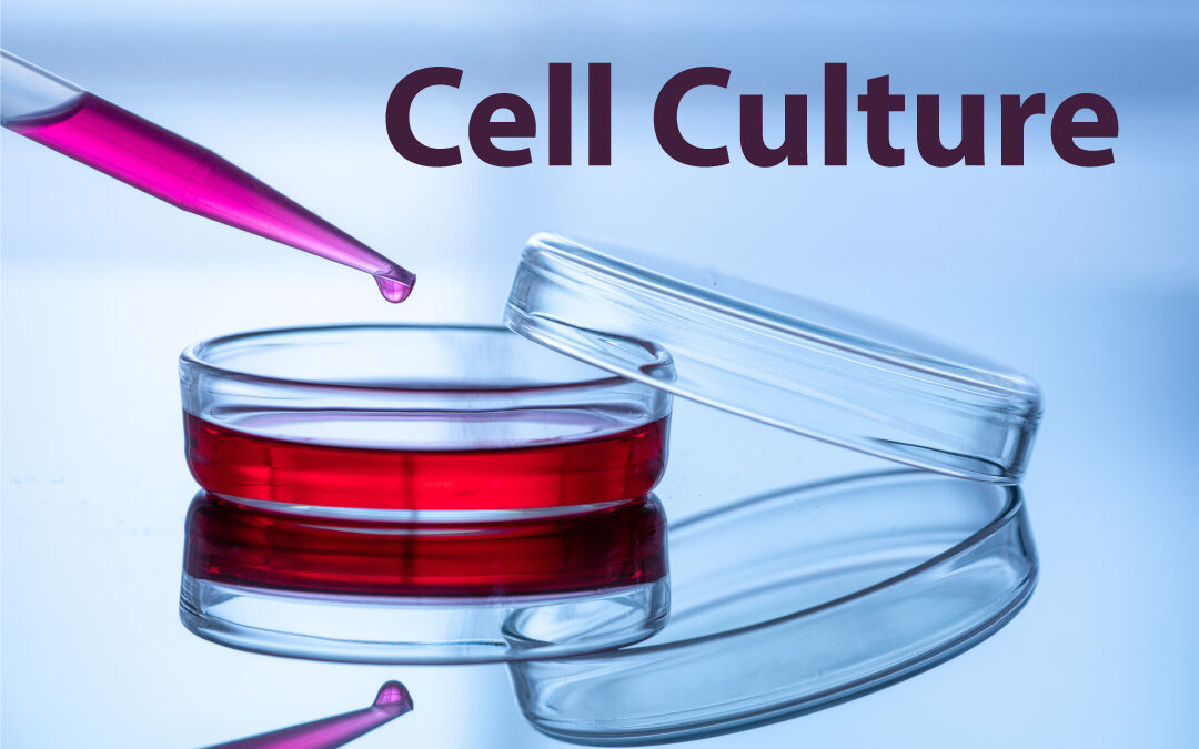
What is cell culture?
Cell culture refers to the process of growing and maintaining cells outside their natural environment in a controlled laboratory setting. This technique allows researchers to study cellular behavior, function, and interactions under conditions that closely replicate those found within the human body.
A brief history of cell culture
To understand where cell culture has come from, we must go back to 1665, when Robert Hooke, an English scientist, first coined the term “cell” while observing a slice of cork under his microscope. He noticed small chambers, which he named “cells”, as they reminded him of small rooms, describing them as the fundamental units of life.
In the early 1900s, American biologist Ross G. Harrison demonstrated the viability of growing animal tissues outside the organism using frog embryos. This laid the foundation for modern tissue culture theory and techniques.
Building upon Harrison’s work, French surgeon Alexis Carrel made significant strides in cell culture during the early 20th century. His innovative techniques, including the use of specialized culture vessels (Carrel flasks) and nutrient-rich media, allowed for long-term cell cultivation and introduced the concept of “continuous” cell lines. Around the same time, German embryologist Wilhelm Roux contributed to the study of embryo development through cell culture experiments, further advancing biological research.
The 1940s saw the introduction of antibiotics to cell culture media, revolutionizing the field by preventing bacterial and fungal contamination. Before this, maintaining sterile cultures was a significant challenge, as contaminants could quickly compromise the integrity of the cultures. Penicillin and streptomycin were among the first antibiotics used, effectively targeting bacterial cell walls and preventing contamination.
Unlike primary cells, which have a limited lifespan, continuous cell lines are immortalized, meaning they can undergo unlimited divisions without losing their genetic profile or characteristics. This makes them essential for studying cellular processes, disease mechanisms, drug screening, and producing biological compounds.
HeLa cells and modern advances
No discussion of continuous cell lines would be complete without mentioning HeLa cells. In 1951, George and Margaret Gey harvested cervical cancer cells from Henrietta Lacks, a poor African American woman, without her knowledge or consent. These cells, named HeLa after her, became the first immortal human cell line, capable of dividing indefinitely in a lab environment. This discovery revolutionized biomedical research, leading to significant breakthroughs such as the development of the polio and HPV vaccines, advancements in cancer research, and numerous insights into cell biology.
However, the story of HeLa cells is also a tale of ethical misconduct and exploitation. For decades, Henrietta Lacks and her family were kept in the dark about the use and commercialization of her cells. It was not until the 1970s that they learned of HeLa’s widespread use in research and the profits it garnered, none of which benefited them.
In 2023, Thermo Fisher Scientific reached a confidential settlement with Henrietta Lacks’ family, addressing the unauthorized use of her cells for research over 70 years ago.
HeLa cells have contributed immensely to science, but also highlight critical issues of medical ethics and racial injustice. Today, Henrietta Lacks is rightfully recognized not just for her unwitting contribution to science but also as a symbol of the need for ethical standards in research.
Further advances
In the late 20th century, more advanced cell techniques emerged:
Transfection: Introducing nucleic acids (DNA or RNA) into cells to change gene expression or produce proteins. Methods include chemical transfection, electroporation (using electrical pulses to open pores in the cell membrane), and viral transduction.
Gene editing: Technologies like CRISPR-Cas9 allow precise genome modification, enabling scientists to create knockout cell lines, introduce specific mutations, or insert reporter genes to study gene function and disease mechanisms.
Co-culture systems: Growing multiple cell types together to simulate and study interactions between different cell populations in vivo. For example, epithelial cells co-cultured with fibroblasts to study tumor development.
3D culture: A major recent advancement, allowing cells to grow in three dimensions. This creates a more physiologically relevant environment compared to traditional two-dimensional monolayer cultures, closely mimicking the structure and function of tissues and organs.
Here at Virology Research Services, we offer air-liquid interface (ALI) testing services to provide advanced, physiologically relevant models of respiratory infections, helping researchers achieve better results in preclinical research. Read more here: ALI cell culture blog and watch our video ALI cell culture.
ALI cell culture
The importance of cell culture
The importance of cell culture cannot be understated, as it underpins many breakthroughs in biomedical research:
Drug discovery: Cell culture plays a crucial role in disease modeling, allowing researchers to replicate medical conditions in the lab. By culturing cells derived from animal models or humans, scientists can mimic disease pathologies, study disease progression, and screen potential therapeutic compounds. These models help identify new drugs, assess their efficacy, and evaluate their toxicity before clinical trials.
Vaccine development and viral research: Cultured cells serve as hosts for viruses used in vaccine production. By growing viruses in cell culture, scientists can study their replication, pathogenesis, and immune responses, aiding vaccine and antiviral therapy development. Cell culture-based systems enable rapid vaccine production and scalability, which is particularly useful during pandemics and outbreaks.
Toxicity testing and safety assessment: Cell culture is essential for evaluating the toxicity of drugs, chemicals, or products. Scientists assess the effects on cell growth, replication, function, and viability, which is crucial for the regulatory testing of new drugs and products.
The role of cell culture in modern biomedical research
Cell culture has transformed – and become a fundamental part of – biomedical research. Its versatility and scalability cater to diverse needs: investigating cellular mechanisms, modeling diseases, exploring novel therapies, and advancing medicine. As technology advances further, cell culture will continue to be at the forefront of drug and diagnostic breakthroughs, opening avenues for discoveries that will transform our health and lives for the better.
If we’ve gone from Harrison to mini-brains in 100 years, and the rate of advance is accelerating, where might we be 10 or 20 years from now?
How can we help?
For further reading, explore our blog articles on advanced cell culture techniques and how we use these methods to study various diseases and develop new therapies. If you need any virus testing services or consultancy, reach out to our team, and an experienced virologist will get back to you right away.
Blog by Paul Griffin
Edited by Reckon Better – Scientific Communication Services