Imagine studying a city from a flat map – you’re missing the depth, the real interactions. Now imagine walking through its streets. That’s the kind of impact 3D cell culture is making for in vitro cell biology.
The Origins of 3D Cell Culture
For decades, 2D cell cultures on flat surfaces like Petri dishes were the standard, but they failed to mimic the complex interactions of real tissues. In the 1980s, scientists began using scaffolds like collagen to create 3D structures, leading to advanced techniques like spheroids, organoids, and bio-printed tissues that better model real cell behavior and disease.
Applications of 3D Cell Culture
3D cell culture has transformed numerous areas of scientific research, including:
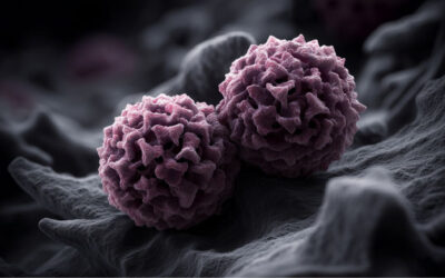
- Cancer Research: Tumor spheroids in 3D cultures replicate the environment of human tumors more closely than 2D models. This allows researchers to better understand cancer behavior, treatment resistance, and the processes that drive metastasis.
- Drug Discovery and Testing: Because 3D cultures simulate human tissues more accurately, they provide a more reliable model for testing drug efficacy and safety, reducing the need for animal testing.
- Regenerative Medicine: Techniques like 3D bio-printing allow scientists to create tissue constructs for repairing damaged organs, paving the way for personalized treatments for conditions such as heart disease.
- Stem Cell Research: 3D environments enhance the maturation and differentiation of stem cells into specialized tissue types, improving the potential for regenerative therapies.
- Disease Modeling: Patient-specific organoids grown in 3D cultures can replicate the structure and pathology of diseased organs, offering new ways to test personalized treatments for conditions such as liver disease or cystic fibrosis.
Virology Applications of 3D Cell Culture
In virology, 3D cell culture has opened new doors for understanding viral infections and developing antiviral therapies. Unlike traditional models, 3D cultures provide a realistic representation of how viruses interact with human tissues, giving researchers better insights into disease progression and potential treatments.
Respiratory Virus Modeling: By simulating the human respiratory tract, 3D cultures have been crucial in studying viruses like influenza and SARS-CoV-2 [for reviews, see Shah et al. (2023) and Ahmed et al. (2023)]. These models help researchers understand viral entry mechanisms, replication, and the effectiveness of potential treatments under conditions that closely mimic human physiology.
For example, studying influenza A can be tricky because traditional 2D cell cultures don’t replicate the complex structure of our lungs. To address this, at Oklahoma State University, Bhowmick et al. (2018) created a 3D lung model using human small airway epithelial cells grown on chitosan-collagen scaffolds at an air-liquid interface, which better mimics the real thing. Their new 3D model not only showed improved cell viability and structure but also provided deeper insights into how lungs respond to influenza infections.
HIV Research: 3D cultures have advanced our understanding of HIV by replicating lymphoid tissues where the virus persists. This allows researchers to observe how HIV reservoirs form and maintain themselves, even under antiretroviral therapy, providing critical insights for developing strategies to eradicate the virus.
For example, HIV can hide out in the brain, leading to HIV-associated neurocognitive disorders (HAND), but studying this has been tough because we didn’t have lab models that truly reflect the brain’s complexity. Now, scientists are using 3D brain organoids made from induced pluripotent stem cells – with microglia included – to better mimic human brain tissue. These innovative models are helping us understand how HIV affects the brain and are opening doors to developing new treatments (Wei et al., 2023).
For more, see this excellent review: 3D human tissue models and microphysiological systems for HIV and related comorbidities
High-Throughput Screening for Antivirals: The adoption of 3D cell culture in antiviral drug screening has improved the speed and accuracy of identifying potential treatments. These models, which closely mimic human organs, are now used to rapidly test the efficacy and toxicity of antiviral compounds. For a review, see Booij et al. (2019).
Advantages Over Traditional 2D Cultures
3D cell culture offers several important advantages over traditional 2D models:
- Physiological Relevance: The 3D structure more closely mimics the natural environment of cells, leading to more accurate predictions of cellular behavior and drug responses.
- Improved Cell-Cell and Cell-Environment Interactions: Cells in 3D cultures develop more naturally, interacting with their surroundings in ways that closely resemble in vivo
- Better Predictive Power: 3D models offer more reliable data for drug testing and disease modeling, bridging the gap between lab research and real-world clinical outcomes.
- Reduced Animal Use: By providing more accurate models, 3D cultures reduce the need for animal testing, helping meet ethical standards and reducing costs.
Challenges of 3D Cell Culture
Despite its advantages, 3D cell culture still faces challenges:
- Complexity and Cost: 3D cultures require specialized materials, equipment, and expertise, making them more complex and expensive than 2D models.
- Reproducibility: Variability in scaffold materials, cell types, and operator techniques can make it difficult to achieve consistent results across experiments.
- Scalability: While advancements are being made, scaling up 3D cultures for high-throughput screening or large-scale tissue engineering remains a challenge.
- Technical Difficulties: Monitoring and analyzing 3D cultures is more complex than in 2D systems, requiring advanced imaging techniques and data analysis tools.
Conclusion
3D cell culture techniques are rapidly becoming an essential tool in biomedical research, offering more accurate models for studying diseases and testing new treatments. With ongoing innovations, the potential applications of 3D cell culture are vast, and we are only beginning to scratch the surface of what this technology can achieve.
Can We Help?
At Virology Research Services, we specialize in 3D air-liquid interface testing services that provide advanced, physiologically relevant models for respiratory infection research. Learn more about how we can enhance your research outcomes here (Air-liquid interface testing services) and watch our video ALI cell culture. Contact our team and one of our scientists will get back to you.
ALI Cell Culture Video
Blog by Paul Griffin
Edited by Reckon Better – Scientific Communication Services