
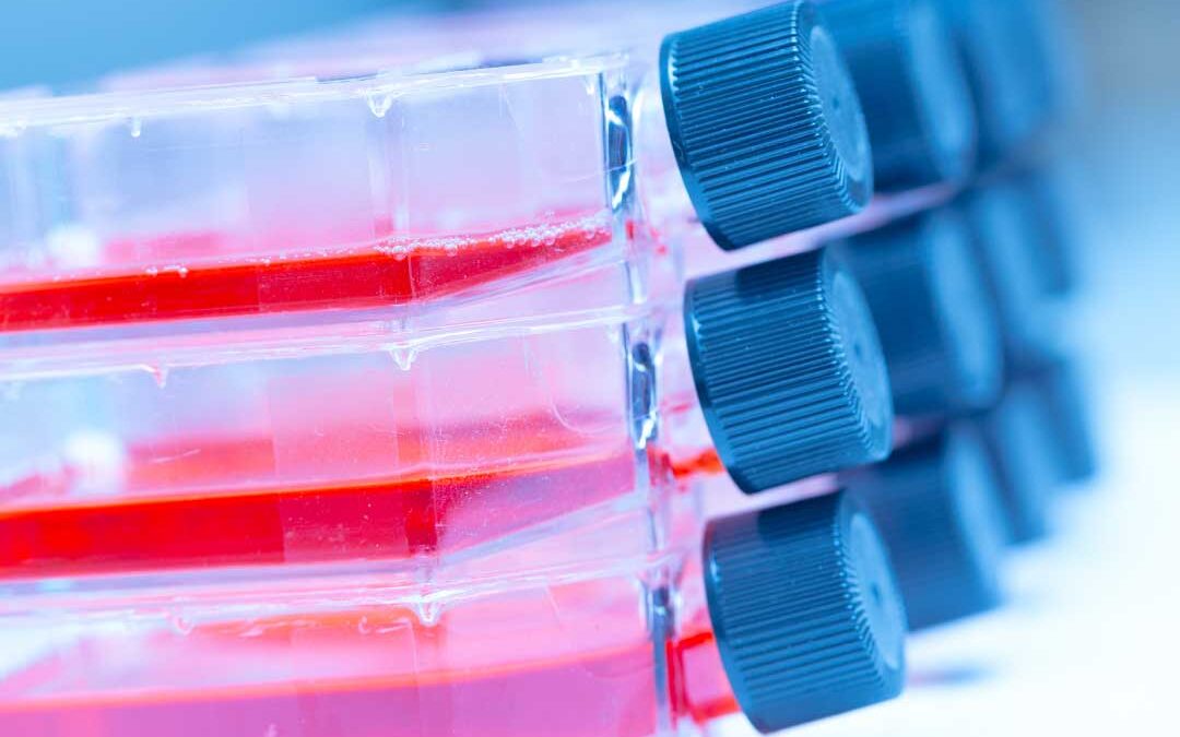
Discussions on antiviral testing often focus on viral strains, compound administration, and assay parameters. Yet one of the most important decisions has already been made behind the scenes: selecting the most appropriate in vitro model.
At VRS, our standard antiviral assays are built around models selected on factors such as viral permissivity, assay robustness, and biological relevance. These models have been carefully established to provide reliable and meaningful data for specific virus systems.
Understanding why a particular model is used, and what information it can or cannot provide, is an important part of interpreting antiviral data.
The Advantage of Simplifying
No in vitro system perfectly replicates the complexity of an infection in a human host, but this is not necessarily a bad thing. A cell culture model removes many layers of biological complexity: multicellular immune responses, tissue organisation, pharmacodynamics, clearance mechanisms, and many other factors that influence viral infection in vivo.
This is certainly a limitation, but the same simplification is also what makes in vitro models so valuable. By reducing complexity, scientists can investigate specific questions in a controlled and reproducible environment.
The aim is therefore to select a model that suits the scientific question being asked and the stage of development, rather than to find a “perfect” one.
What Drives the Choice?
The virus
Very often in virology, the options are limited. Many viruses replicate only in specific cell lines that are neither physiological nor derived from the relevant host tissue, while others require highly specialised 3D systems that do not lend themselves well to robust or multi-sample applications.
Cells with even a partially intact interferon response (the early-warning signalling that switches on a cell’s antiviral defences) can present a significant barrier to in vitro infection. This is one reason why several of the most widely used propagation lines are interferon-deficient. Vero cells lack the entire type I interferon gene cluster through a homozygous deletion on chromosome 12, which is part of why they support such a broad range of viruses. As a result, the most physiological model, such as primary cells with the correct tropism and an intact innate immune response, may produce little or no detectable infection, or require unrealistically high viral inocula to infect.
In many cases, therefore, the biology of the virus makes the first decision for us.
Balancing relevance and reproducibility
One of the central challenges in virology research is balancing biological relevance with experimental practicality.
Complex and physiologically relevant in vitro models tend to require expensive set-ups, specialised reagents, and extensive hands-on time. They are often difficult to grow in large numbers or on standard laboratory plasticware, making them impractical for assays in which multiple compounds, concentrations, controls, and replicates need to be tested. Complexity also tends to come at the expense of reproducibility, with greater variability between donors and batches, and the absence of standardised protocols makes results harder to compare across laboratories.
For these reasons, a model used for early screening or comparative IC50 and TC50 determination may need to prioritise robustness (independent of donor-specific variability), scalability (to accommodate multiple replicates and controls, which at this stage are just as important as the test compounds), and reproducibility.
Developing a system in which a virus and a cell interact consistently, experiment after experiment, is already a significant challenge. Introducing additional variables during the early stages of drug discovery, when the primary focus is identifying promising candidates, can be counterproductive.
By contrast, a model used for later-stage characterisation may place greater emphasis on biological relevance and physiological similarity. Once a compound has demonstrated activity against multiple influenza strains in a simpler cell line system such as MDCK-II or A549, and its IC50 has been established, researchers may wish to evaluate its performance in an air-liquid interface (ALI) model. Being closer to the respiratory epithelium, this system can provide additional information on efficacy and cytotoxicity in the presence of mucus, multiple epithelial cell populations, and an interferon response.
Neither approach is inherently better. The important question is whether the model is proportionate and appropriate for the decision the study needs to support. Good experimental design recognises these trade-offs: a robust assay is not necessarily the most complex one, and a physiologically relevant model is not automatically the most informative at every stage of development.
The test article
The test article determines the scientific question being asked, and this in turn influences which assay should be performed and which cells should be used.
A neutralisation assay designed to assess the ability of an antibody or serum to block infection, or an assay intended to determine the virucidal properties of a disinfectant, typically requires the most permissive cells available for the virus of interest. The objective is to detect as many infectious particles as possible that remain capable of initiating infection following treatment. A poorly permissive cell line, even if more physiological, would not adequately answer these questions if infection is already limited before treatment.
An antiviral compound, particularly one targeting host pathways or specific virus-host interactions, may instead benefit from a more physiologically relevant model. This does not necessarily mean primary cells from the outset, but where possible it may involve cell lines derived from the relevant tissue (for example, lung or liver) or cell type (such as epithelial cells or fibroblasts) infected in vivo.
The assay endpoint
The assay readout can further influence the choice of model.
If antiviral activity is measured through cytopathic effect (CPE), assessing whether the treatment prevents virus-induced cell death, this immediately limits the choice of cell line. Some cells produce a rapid and well-defined cytopathic effect within a timeframe compatible with the assay, while others do not. Similarly, plaque or focus-forming assays require cells capable of producing a uniform and stable monolayer.
Different readouts also answer different biological questions. Measuring infectious virus, viral genome levels, viral protein expression, or changes in cell viability each provides different information and may require different experimental systems for optimal performance.
Looking beyond the first experiment
A well-designed study should answer the immediate question and help guide future decisions. Does the assay provide the information needed to understand the potential of a candidate? Are the results sufficiently robust to inform the next stage of development? Will the data support progression to more advanced models?
Understanding the Limitations
While antiviral efficacy tends to be relatively conserved, particularly for direct-acting antivirals, negative results often require much more careful interpretation.
Take cytotoxicity as an example. Cytotoxicity must always be measured alongside antiviral activity to determine whether reduced viral replication is genuinely due to antiviral activity, or simply reflects the fact that the treatment has killed the host cells.
From the perspective of an antiviral assay, therefore, the primary role of cytotoxicity measurements is to determine whether the antiviral data can be interpreted reliably. Any further conclusion needs to be assessed more deeply. A compound that appears cytotoxic in cultured cells is not necessarily toxic in a whole organism, where cells are supported by tissues, repair mechanisms, metabolism, and multiple clearance systems. Equally, some compounds appear relatively non-toxic in vitro but prove highly toxic in vivo.
Negative antiviral results can also have many explanations. A compound may fail to show activity because the pathway it targets is redundant in the chosen cell line, which raises the important question of whether that pathway is equally redundant in vivo. A prodrug may not be correctly metabolised by the cells being used, preventing activation of the compound. In other cases, we have observed compounds that are rapidly pumped out of cells by membrane transporters, resulting in poor intracellular accumulation and apparently weak antiviral activity. This effect is well documented for P-glycoprotein, where efflux activity lowers intracellular drug concentrations and measured potency shifts with the transporter expression of the cell line used.
Whenever we obtain a result, it is therefore important to interpret it in the context of the model that generated it. Beyond identifying experimental artefacts, understanding the limitations of the model can provide valuable clues about what may happen later in more complex biological systems.
There Is No Perfect Model: Only the Right Model for the Question
Every in vitro system has strengths and limitations. Recognising those limitations is part of good science.
At VRS, our validated assay platforms remove much of this complexity for clients by providing established systems designed around specific virus biology and assay readouts. Sometimes the choice is dictated by the virus itself, sometimes by the practical requirements of the assay, and sometimes there is the opportunity to move progressively towards more physiologically relevant systems as compounds advance through development.
RSV provides a good example. Antiviral screening can be performed in HEp-2 cells, a highly permissive and well-characterised model for RSV infection. The choice matters. HEp-2 cells support markedly greater RSV replication than A549 cells, which mount a more potent interferon-driven antiviral response. We have also extended the assay to A549 lung epithelial cells to provide a more physiologically relevant system, and more recently into primary bronchial epithelial cells cultured at an air-liquid interface. Each step increases biological relevance while answering different scientific questions, allowing compounds to be evaluated in progressively more representative models as they advance through development.
Understanding the rationale behind model selection helps researchers interpret their data and make better decisions throughout the development process. The value of an experiment comes from choosing the right simplification to answer the right question.
How VRS Can Help
Our team can help you select and design the right model for the stage your programme has reached, from early antiviral drug screening in highly permissive cell lines through to more physiologically relevant systems. Get in touch to discuss the right approach for your compound.
References
- Sakuma C, et al. Poliovirus-nonsusceptible Vero cell line for the World Health Organization global action plan. Scientific Reports. 2021;11:6746. doi: 10.1038/s41598-021-86050-3
- Rajan A, et al. Multiple respiratory syncytial virus (RSV) strains infecting HEp-2 and A549 cells reveal cell line-dependent differences in resistance to RSV infection. Journal of Virology. 2022;96(7):e01904-21. doi: 10.1128/jvi.01904-21
- Ekanger CT, et al. Comparison of air-liquid interface transwell and airway organoid models for human respiratory virus infection studies. Frontiers in Immunology. 2025;16:1532144. doi: 10.3389/fimmu.2025.1532144
- Translatability of in vitro inhibition potency to in vivo P-glycoprotein mediated drug interaction risk. Journal of Pharmaceutical Sciences. 2023. Available here